The research team of Frontiers Science Center for Nano-Optoelectronics has made significant progress in the research of New Single-Lens Light-Sheet Imaging Technology
Information: Frontiers Science Center for Nano-Optoelectronics
In traditional fluorescence microscopy, two vertically arranged objective lenses are generally used for light excitation and signal collection. The design of such a dual objective optical microscope system needs to consider the matching relationship between the numerical aperture of the objective and its working distance, which limits its utilization of large numerical aperture objectives and results in limited spatial resolution; At the same time, the vertical placement of this dual objective optical path structure also makes the adjustment of the imaging system complex and inefficient, so the development of single-lens light-sheet fluorescence microscopy systems is particularly important.
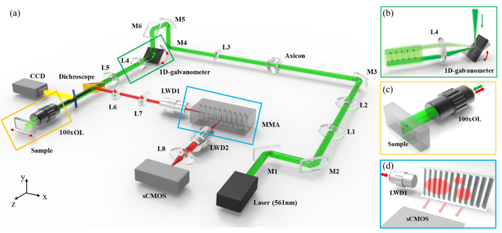
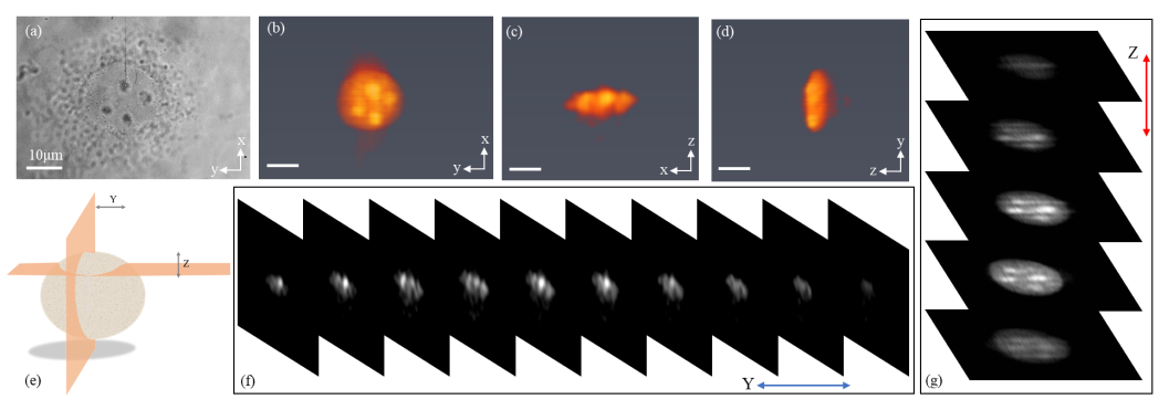
Based on the above research environment, the team has invented a single objective fluorescent imaging system that combines a micro-mirror array. As shown in Figure 1, this micro-mirror array is composed of a series of evenly spaced flat micro mirrors, each of which maintains the same tilt angle. After the excitation light sheet excites the sample in a plane, the fluorescence information generated will be collected by the same objective lens and transformed into a two-dimensional plane perpendicular to the optical axis through the array of micro mirrors, which will be received by subsequent imaging units. The single-lens fluorescence imaging technology successfully achieved imaging of 10um/2um fluorescence microspheres and live cells, as shown in Figure 2.

Fig.1 Schematic diagram of the Single-Lens Light-Sheet Fluorescence Microscopy based on micro-mirror array

Fig.2 Imaging results of COS-7 cells
This new single objective in light sheet imaging method innovatively improves the structure of traditional optical fluorescence imaging and makes it easier for ordinary fluorescence microscopy systems to upgrade to optical imaging. It has broad application prospects in the field of high spatiotemporal resolution imaging.
The co-first author of the paper is Yanhui CAI, PhD, School of Physics, Peking University, 2016; Yizhu Chen, PhD, Pennsylvania State University, 2016; corresponding author is Kebin Shi, Professor, Peking University.
The above research work was supported by the Major Research Program of the National Natural Science Foundation of China, the National Biomedical Imaging Center and the Major Basic and Applied Basic Research Project of Guangdong Province.


