


Peking University Leads Preparations for VITAL Initiative
Source: Peking University Media Center & National Biomedical Imaging Center (NBIC), Peking University
On the evening of August 26, 2026, at the opening ceremony of the HICOOL 2026 Global Entrepreneur Summit, the Panoramic Digital Life International Big-Science Program (VITAL Initiative)—led by Peking University with Academician Cheng Heping as Chief Scientist—was formally launched into its preparatory phase. The initiative will be based at the Huairou Comprehensive National Science Center.
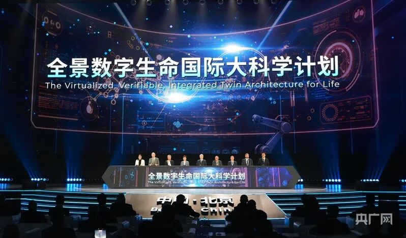
On the evening of August 26, 2026, the VITAL Initiative was formally launched into its preparatory phase at the opening ceremony of the HICOOL 2026 Global Entrepreneur Summit (Photo by China National Radio, provided by the Publicity Department of the Huairou District Committee)
In Huairou Science City, the Multimodal Trans-Scale Biomedical Imaging Facility, constructed under Peking University’s leadership, is already operational. It enables whole-body imaging in a single bed position with a full-body positron emission tomography/computed tomography (PET/CT) system; freely moving mice can wear a 2.6-gram miniaturized microscope that allows researchers to observe the dynamics of deep-brain cells; and cryo-electron microscopy extends the view to the fine structures of proteins and other biomolecules.
These vastly different observational scales are integrated on a single facility platform, which serves as a critical foundation for the VITAL initiative.
Digital life requires not only static three-dimensional morphological depiction but also the revelation of the processes and principles of life activities. A digital cell must show how its internal components interact and how structure and function change over time; a digital organ must reflect how different cells coordinate and how local changes affect the whole.
Whether a model can be used for research depends on its ability to withstand testing in real application scenarios.Building such models first requires recording life states as realistically and continuously as possible, along with how those states change over time and under perturbations. The human body, organs, cells, and molecules exist at different scales and are constantly changing—where do the necessary data come from? This facility in Huairou Science City is providing the conditions to answer that question.
Seeing Life: One Facility Spanning from Molecules to the Human Body
On March 21, 2025, after 12 years of planning, construction, and trial operation, the Multimodal Trans-Scale Biomedical Imaging Facility passed national acceptance. Peking University served as the legal entity and lead construction unit, jointly building the facility with the Institute of Biophysics of the Chinese Academy of Sciences and other partners. The National Biomedical Imaging Center (NBIC) was established on the basis of the facility to undertake open operation, technology development, and research organization.
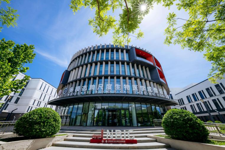
Exterior view of the Multimodal Trans-Scale Biomedical Imaging Facility
Viewed from above, six white buildings are arranged along a golden spiral. The circular building resembles a cell—or an “eye” gazing at life. From architectural layout to interior spaces, the facility is organized around different scales of life observation and experimental conditions.
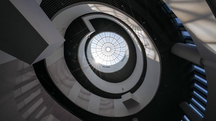
“Eye of Life” design inside the facility
The Sample Preparation and Model Animal Center supports experiments with biological samples, cells, and animal models. The Multimodal Medical Imaging Device primarily observes structural and functional imaging of the tissues and organs of complete living bodies, from small rodents to human beings; and the Multimodal High-Resolution Molecular Imaging Device reveals subcellular and molecular structures. Data generated by the different devices can enter the Full-Scale Image Data Integration System for storage, registration, computation, and analysis. Researchers select one or more platforms according to their scientific questions so that observations at different scales form connections.
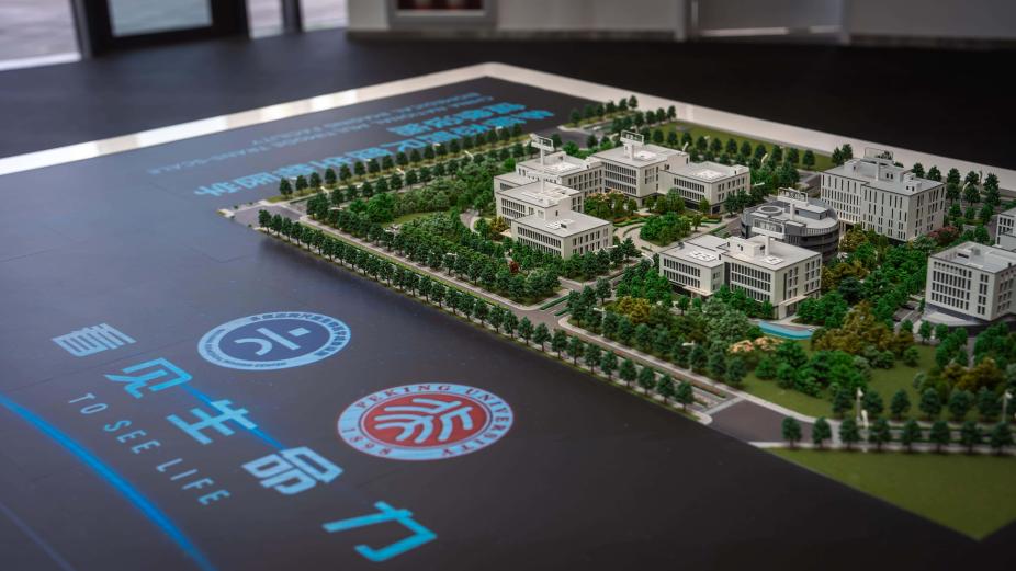
Scale model of the National Biomedical Imaging Center, Peking University
The facility integrates optical, acoustic, electrical, magnetic, radionuclide, and electron imaging modalities to record different aspects of life activities. Some are suited to anatomical structure, others to metabolism and function, neural electromagnetic activity, or the fine structure of molecules. Multiple imaging approaches cross-reference one another, helping overcome the limitations of any single perspective.
The spatial observation range extends from angstroms to meters, connecting molecules, cells, tissues, organs, and whole organisms. After seeing life’s landscapes at different scales, researchers continue to ask: How does a minute molecular event alter cell behavior? How do changes in a group of cells produce phenotypes at the organ or organism level?
Decoding Life: Seeking Connections Across Scales
Decoding life requires linking the whole with the part, structure with function, and static images with dynamic processes. The facility provides a path of progressive deepening and iterative verification: discover anomalies at the human-body and organ level, track changes in the living state, seek mechanistic clues at the molecular level, and then connect evidence across scales.
Macro scale – Finding anomalies in the human body and organs
In disease research, imaging at the human-body and organ levels often supplies the initial clues. Where an anomaly occurs, which tissues it affects, and whether it is linked to other organs all require judgment from the whole.
The facility’s full-body PET/CT features a 2-meter axial field of view and enables single-bed whole-body imaging. PET uses tracers to track metabolic and molecular activity in the body, while CT provides the corresponding anatomical location. Combining the two allows researchers to observe how tracers distribute, where they accumulate in organs, and how that distribution changes over time. The system currently primarily serves scientific research.
Magnetic resonance imaging, magnetoencephalography, and other equipment supply information on structure, blood flow, function, and neural electromagnetic activity. Different imaging modalities complement one another, enabling researchers to identify regions and processes that warrant further tracking amid complex whole-organism activity.
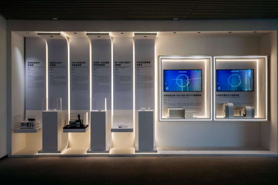
Introduction to the facility’s highlight equipment in the exhibition hall
Meso scale – Tracking cellular changes in vivo
After identifying clues from whole-organism imaging, research must enter tissues and cells to observe how changes occur. Fixed samples capture a single moment; live imaging can record how organelles communicates, when neurons are activated, and how cells transmit signals. The Multimodal Live-Cell Imaging Device covers subcellular, single-cell, tissue, and small model-animal levels and can observe structural and functional changes under near-physiological conditions.
Here, mice can freely move while wearing a miniaturized two-photon microscope. The new-generation multicolor miniaturized two-photon microscope released in 2025 weighs 2.6 grams and uses different fluorescent labels to record the dynamics of different cells or subcellular structures in the deep brain of freely behaving mice. Researchers can thereby correlate neural signals with behaviors such as movement, feeding, and sleep.
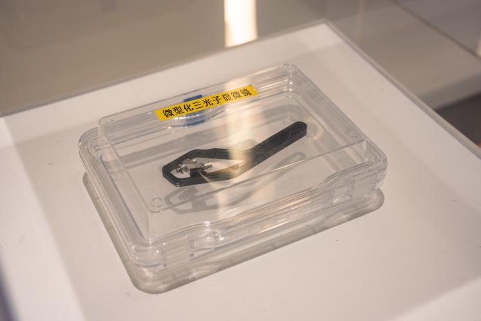
Miniature multiphoton microscope
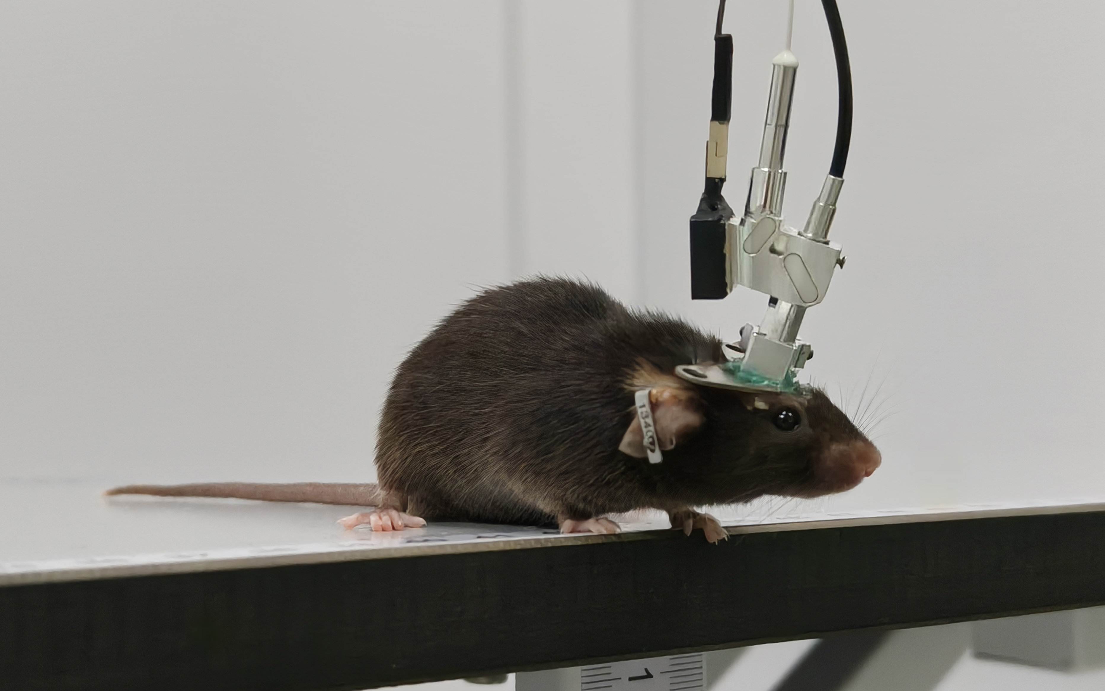
Multicolor miniature two-photon microscope
Stable, precise observation also depends on the building’s engineering design. The live-cell imaging platform houses more than 30 ultra-precision instruments; even slight ground vibration can shift images. To isolate disturbances, the entire floor of the equipment rooms is lifted by compressed air, forming a large air-bearing platform. Precision instruments, building structure, environmental control, sample preparation, and experimental workflows together constitute the facility’s research capability.
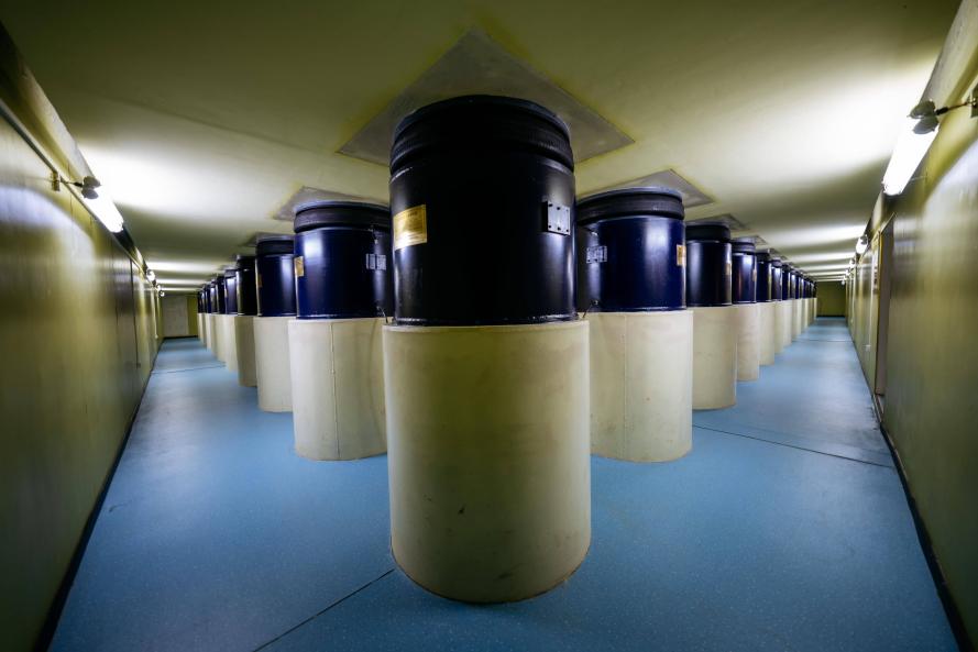
Large air-bearing anti-vibration platform
Micro scale – Seeking mechanistic clues in molecular structure
Looking deeper, the question shifts from “which cells have changed” to “which molecular events may have caused those changes.”
The Multimodal High-Resolution Molecular Imaging Device can observe proteins, viruses, DNA, and the ultrastructure inside cells. Cryo-electron microscopy images rapidly frozen biological samples and, under suitable conditions, yields structural information approaching atomic resolution. Researchers can analyze how proteins fold, how different molecules bind, and where drugs may act. Ultrafast electron microscopy and related techniques add the temporal dimension, helping observe rapid processes.
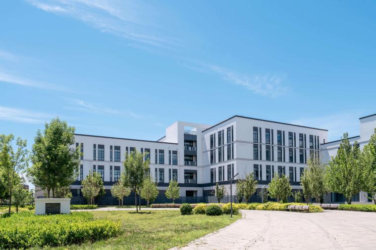
Exterior of the Multimodal High-Resolution Molecular Imaging Platform
Molecular structures supply mechanistic clues. Researchers must then return to cells, tissues, and whole organisms to test whether these changes show stable associations with the observed functional anomalies. A scientific question thus completes a round trip: from whole-organism phenotype down to microscopic structure, then back up with larger-scale experiments to test the initial explanation.
Integration – Connecting the Part and the Whole through Cross-Validation
Images generated by different devices differ in resolution, coordinate systems, sampling times, and imaging principles. The Full-Scale Image Data Integration System handles data storage, computation, registration, and analysis. To uncover deeper connections, experiments must be designed around a common research question, allowing observations made at different scales in the same specimen, matched specimens, or animal models to be compared and cross-validated.
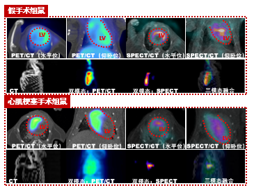
Application of multimodal image analysis in research
A concrete example comes from the research of Professor Chen Liangyi’s team at Peking University’s College of Future Technology on insulin secretion.
After a rise in glucose, the pancreatic islet first rapidly releases a batch of insulin, then enters a longer-lasting second phase of secretion. Previously, this process was mainly explained through vesicles and molecular mechanisms inside individual β-cells. Researchers simultaneously observed vesicle fusion, single β-cell activity, and the secretory response of the intact islet. Under the conditions of that study, β-cells showing clear secretory activity accounted for about 40 percent; temporal synchrony and asynchrony among different cells together shaped the biphasic secretion. In obese model mice, the number and secretory patterns of these cells were abnormal.
The study thus presents biphasic insulin secretion as a tissue-level function formed by the coordination of different β-cells. Information at the vesicle level cannot explain the secretory rhythm of the whole islet, while tissue-level observation cannot determine which cells account for the differences. Only when vesicle exocytosis, single β-cell secretion, and whole-islet function are linked across three scales do the previously scattered phenomena form a relatively complete chain of mechanistic evidence.
This work was completed before the facility passed national acceptance and can be regarded as an early sample of the cross-scale research approach. The facility provides the conditions to extend such observation to more scales, imaging modalities, and life-science questions, and through the data system converts images into information that can be compared, computed, and modeled.
Modeling Life: Subjecting Digital Models to Experimental Validation
Moving from seeing and decoding life toward digital reconstruction requires the joint support of multimodal trans-scale imaging, spatial omics, and verifiable computational models. The imaging facility undertakes one foundational task: continuously acquiring structural and functional data of real life and providing experimental validation conditions for digital models.
In the future, when a model predicts that a certain class of molecular change may affect cell and organ function, researchers can design cross-scale imaging experiments accordingly and then use the experimental results to correct the model. Data support model building; models raise new questions; experiments test model predictions—forming an iterative research process.
Peking University has already carried out years of scientific research and organizational preparation around digital life. In April 2025, PKU launched the first batch of major digital-life projects on the basis of the imaging facility; model directions such as digital C. elegans, digital kidney, and digital islet have advanced step by step. In April 2026, Peking University signed a memorandum of cooperation with ten institutions including relevant institutes of the Chinese Academy of Sciences, Tsinghua University, and Wuhan University, further advancing domestic collaboration on the VITAL Initiative. In August 2026, the HICOOL 2026 Global Entrepreneur Summit further initiated preparatory work on international organizational mechanisms, common standards, a transnational validation network, and the Huairou hosting system.
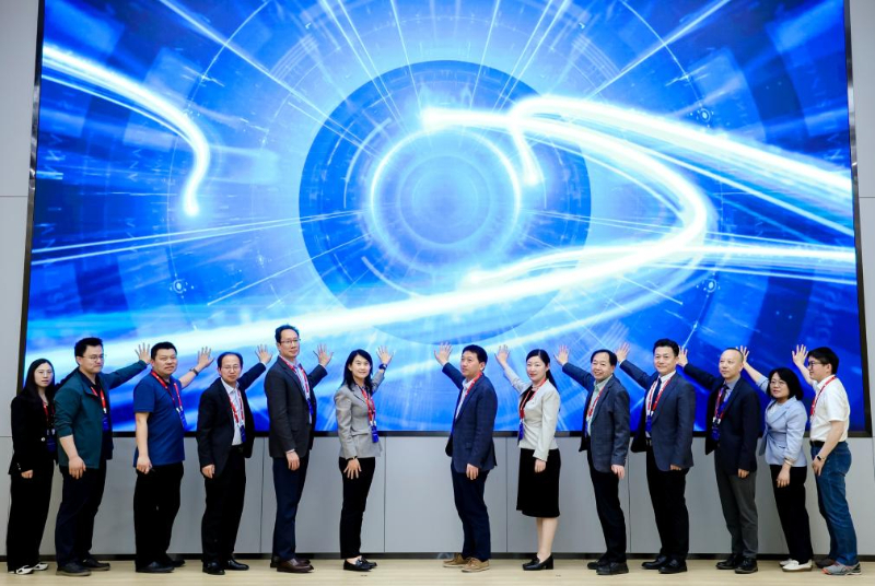
In April 2025, Peking University launched the first batch of major “Digital Life” projects based on the imaging facility
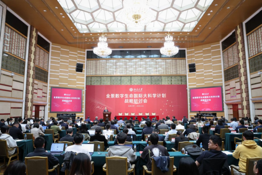
On April 21, 2026, the Strategic Seminar on the VITAL Initiative was held at Peking University
The facility is also accumulating data and operational experience through open running. To date it has served more than 400 research groups from over 30 institutions, with instrument usage exceeding 40,000 hours; nearly 100 core imaging instruments are open for reservation by research institutions and enterprise users. Researchers from different disciplines use the equipment and conduct experiments here, creating conditions for reusable data and methodological standards.
For the VITAL Initiative, the value of the imaging facility lies not only in producing more and clearer images, but in allowing the predictions of digital models to return to real living systems for testing. Atlases tell researchers “what is there and where”; models further answer “which states and rules determine the next change.” Only models that can withstand prospective perturbation testing can become true digital twins. From molecules to the human body, from a rapid reaction to the long-term evolution of disease, Peking University is working to connect previously dispersed observational capabilities and to place “seeing life” and “validating life” on the same scientific chain.