Chen Zhixing's Team and Zou Peng's Team collaborated to develop a milder far red membrane voltage indicator
Information source: Professor Chen Zhixing's team
On August 17, 2023, the research group of Chen Zhixing, College of Future Technology, Peking University, National Biomedical Imaging Science Center, and the research group of Zou Peng, School of Chemical and Molecular Engineering, Peking University, were published in Proc.Natl.Acad.Sci.U.S.A. The journal published the results of the latest collaborative study "Orange/far-red hybrid voltage indicators with reduced phototoxicity enable reliable long-term imaging in online. neurons and cardiomyocytes ", they synthesized cyclocettaene (COT) modified cyanine dyes with good photostability, and combined them with HVI (hybrid voltage indicator) with excellent sensitivity to upgrade them into a new generation of low-phototoxicity probes. Among them, the orange fluorescent probe HVI-COT-Cy3 can record the membrane potential of neurons for 30min, and can effectively identify the effects of drugs on the electrophysiology of neurons and cardiomyocytes. In addition, the far red probe HVI-COT-Cy5 can be combined with the green fluorescence probe to realize the parallel detection of nerve membrane potential and calcium signal for continuous 15 min.

The key to unlocking the mysteries of the brain is to study the communication mechanism between cells, and the core of understanding the electrical communication between cells is to measure and interpret the electrical signals between cells. Compared with traditional patch-clamp technology, membrane potential imaging has the advantages of non-invasive detection and high detection flux, and has gradually emerged in the detection of spatiotemporal dynamic changes of excitable cell electrical signals. Fluorescent membrane potential probes based on the combination structure of voltage sensitive proteins and small molecular fluorescent dyes show obvious advantages in voltage sensitivity, light stability and cell specificity, which greatly promote the application of optical imaging in detecting neural activity.
However, the excitation light required for fluorescence imaging is inherently phototoxic to living cells, and in order to obtain a sufficiently high signal-to-noise ratio, membrane potential imaging requires a higher excitation light intensity, resulting in more obvious phototoxicity and more rapid photobleaching, which greatly limits the recording duration of membrane potential imaging. For neuroscience researchers and drug developers, whether studying synaptic plasticity, animal behavioral rhythms, or characterizing pharmacological toxicity, continuous or cumulative electrophysiological recordings are often required for a long time, which greatly tests the photostability and phototoxicity of membrane potential probes.
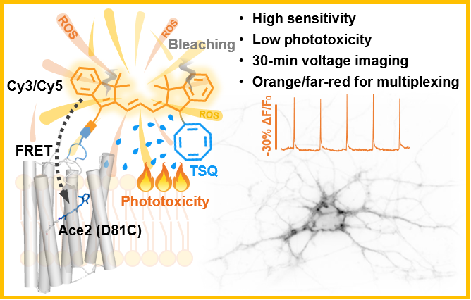
Zou Peng Lab has long developed chemical probes for detecting biological signals, and first proposed the concept of "composite membrane potential probes", which used bioorthogonal chemical labeling strategies to pair fluorescent dyes with rhodopsin proteins, and realized cell membrane potential observation through electrochromic effects (Nat.Chem. 2021, 13, 472-479). However, in their experiments, they found that fluorescent probes based on chemical dyes were difficult to avoid phototoxic damage to neurons. Chen Zhixing's research group has been committed to tailoring chemical molecules according to chemical principles to solve the problem of "technology jam" in biological research. In 2020, Chen Zhiheng's research group published an article (Chem.Sci.2020, 11, 8506-8516), synthesizing a mitochondrial probe using the three-line state hardener cycloctadiene (COT) coupling strategy, demonstrating for the first time the potential of low-phototoxicity probes in the field of super-resolution imaging at the living cell level. Therefore, the two research groups hit it off immediately and launched a joint research on the pain point of phototoxicity in membrane potential imaging. By synthesizing COT-Cy3 and COT-Cy5, COT-modified cyanine dyes, the team developed a new, gentler membrane potential probe, HVI-COT, which significantly improved phototoxicity and light stability of membrane potential imaging (Figure 1).

Figure 1. Concept diagram of low phototoxicity composite membrane potential probe
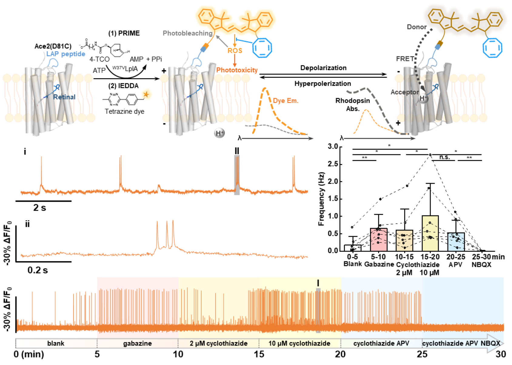
The experimental results show that the sensitivity of HVI-COT-Cy3 (-33.4±0.8% ΔF/F0 per AP) exceeds that of HVI-Cy3, setting a new high for the sensitivity of the orange band membrane potential probe. With excellent photostability and slight phototoxicity, HVI-COT-Cy3 can continuously record the membrane potential of neurons for 30 min, and identify the effects of multiple drugs on the burst frequency of neuron action potential within this time window (Figure 2). It is worth mentioning that the photostability of HVI-COT-Cy3 is more than 40 times that of the similar probe Voltron2525, and the phototoxicity is significantly lower than Voltron2525.

Figure 2. Fluorescent labeling method of HVI probe, principle of membrane potential response, and results of long-term recording of neuronal electrical activity
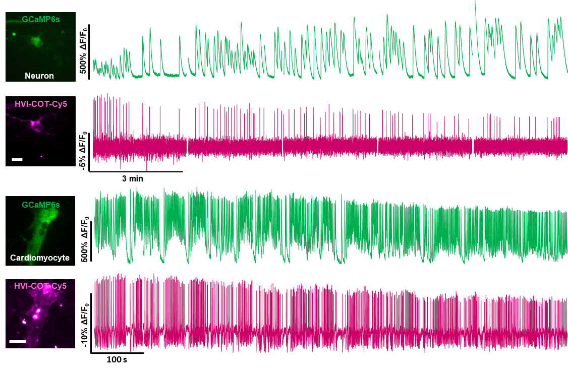
In addition, HVI-COT-Cy5 can be paired with the green fluorescent calcium probe GCaMP6s to perform bicolor imaging in neurons and cardiomyocytes for 15 minutes. Multicolor imaging helps researchers understand the relationships and differences between a variety of dynamically changing physiological signals. In the future, HVI-COT-Cy5 is expected to be combined with green calcium probes to evaluate the neurological or myocardial toxicity of drugs with dual indicators.

Figure 3. HVI-COT-Cy5 was used to record electrocalcium signals of neurons and cardiomyocytes simultaneously with GCaMP6s
In summary, the work developed a milder high-performance fluorescent probe capable of recording electrical signals of neurons and cardiomyocytes for a long time by modifying chemical fluorescent dyes for phototoxic pain points in the field of membrane potential imaging, and realized long-term multicolor imaging and pharmacological detection. In recent years, the depth and breadth of functional membrane potential imaging applications have been expanding, moving towards more diverse research systems, deeper in vivo observation areas, and longer recording duration. The HVI-COT series of probes is expected to become an important tool for researchers to more reliably evaluate the electrophysiological effects of drugs on neurons or cardiomyocytes.
Shuzhang Liu, Ph.D., School of Chemistry and Molecular Engineering, Peking University, and Jing Ling, Ph.D., Institute of Frontier and Interdisciplinary Studies, Peking University, and Peking University - Tsinghua Joint Center for Life Sciences are co-first authors of the paper. Chen Zhixing, a researcher at the National Center for Biomedical Imaging Science and the College of Future Technology at Peking University, and Zou Peng, a researcher at the School of Chemical and Molecular Engineering at Peking University, are co-corresponding authors of the paper. This work was supported by funding from the Beijing Municipal Commission of Science and Technology, the National Natural Science Foundation of China, the Ministry of Science and Technology, the Key Laboratory of Bioorganic and Molecular Engineering of the Ministry of Education, the Beijing National Research Center for Molecular Sciences, the IDG/ McGovern Institute for Brain Sciences of Peking University, and the Peking University Tsinghua Joint Center for Life Sciences.
The original link: https://www.pnas.org/doi/10.1073/pnas.2306950120


