Peng Xi's team develops Multi-resolution analysis enables fidelity-ensured deconvolution for fluorescence microscopy
Source: Peng Xi's Research Team
Fluorescence microscopy serves as a vital tool in live-cell imaging. The introduction of super-resolution microscopy has further breached the diffraction limit of fluorescence microscopes, enabling the observation of finer cellular structures. However, random noise and optical blurring during imaging processes hamper the quality of fluorescence imaging. Moreover, in live-cell imaging, high photon doses adversely affect cell states and lead to photobleaching, increasing the difficulty in obtaining high signal-to-noise ratio (SNR) images. Moreover, insufficient exposure time in ultrafast imaging restricts the acquisition of high SNR images. In summary, key parameters such as imaging speed, duration, and resolution in fluorescence imaging are significantly constrained.
Deconvolution algorithms have gained substantial applications in fluorescence imaging due to their potential to improve imaging duration and spatiotemporal resolution by recovering fluorescent signals with a low photon budget. Although algorithm compensation has demonstrated immense potential in enhancing these crucial aspects of fluorescence imaging, their fidelity, especially in resolution enhancement, remains questionable. Furthermore, developing deconvolution algorithms capable of operating at even lower SNRs could further enhance imaging duration and speed, offering more opportunities for discovering novel biological phenomena.
To address these issues, Professor Xi Peng's team at Peking University has reshaped the noise control model and resolution enhancement mechanism of fluorescence imaging deconvolution algorithms, proposing the Multi-resolution Analysis (MRA) deconvolution algorithm. This method effectively elevates the minimum SNR limit that deconvolution algorithms can recover and overcomes the artifacts produced by traditional deconvolution algorithms during resolution enhancement. This research has been published in eLight under the title "Multi-resolution analysis enables fidelity-ensured deconvolution for fluorescence microscopy."
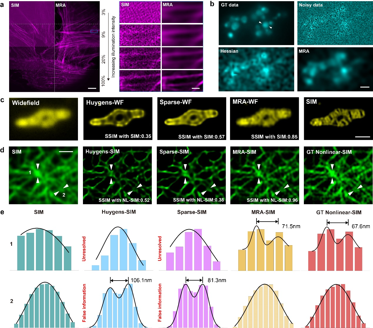
The researchers initially analyzed the shortcomings of conventional variation-based regularization methods including total variation (TV) and Hessian regularization, arguing that while they partially capture the continuous features of fluorescence imaging, they also confuse fluorescent signals with background edges and struggle to extract signals from extremely high noise. Based on the specific labeling and biological structural characteristics of fluorescence imaging, the researchers first proposed two cardinal attributes to differentiate fluorescent signals from noise: (1) high contrast across edges and (2) high continuity along edges. Mathematically, the authors employed multi-scale framelet and curvelet transforms to extract these two key features in fluorescent signals, termed the MRA regularization method.
Regarding resolution enhancement, the researchers initially demonstrated through simulation experiments that the Richardson-Lucy iteration spontaneously generates artifacts due to its statistical estimation characteristics. While iterative inverse filtering based on physical models can perfectly restore blurred images in noise-free conditions, the recovery of high-frequency details requires numerous iterations, which are easily overwhelmed by noise and poorly compatible with variance-based noise control regularization. In contrast, the proposed MRA regularization method, by providing a more accurate fluorescence noise model, effectively controls noise in the inverse filtering iterations while preserving the inferred high-frequency details. Furthermore, the researchers introduced an accelerated FISTA iteration strategy that can perform thousands of constrained inverse filtering iterations within a short computation time to restore high-frequency details.
Through experiments restoring images under various imaging conditions with varying SNRs, the researchers verified that the MRA regularization method could further improve SNR by 2-10 dB compared to variation based regularization methods.

Figure 1. MRA deconvolution achieves superior noise control and fidelity-ensured resolution enhancement. a. Validation of MRA noise control using SIM images with varying SNRs. b. Comparison between MRA and Hessian regularization in continuous frame image processing. c. Images of U2OS cell mitochondria, with deconvolution results from Huygens, Sparse, and MRA, alongside the SR-SIM ground truth. d. Pairs of linear and nonlinear SIM images obtained from the open-source BioSR dataset. Nonlinear SIM serves as the ground truth for deconvolution results from Huygens, Sparse, and MRA on linear SIM images. e. Intensity profiles along the lines indicated by the two white arrows in d.
Using multiple high-low resolution data pairs, including widefield-SIM, linear SIM-nonlinear SIM, confocal-SoRa, and confocal-STED, the researchers verified that MRA deconvolution exhibits significantly superior fidelity compared to deconvolution algorithms using Richardson-Lucy for resolution enhancement, with resolved high-frequency details that almost match those of super-resolution microscopes.
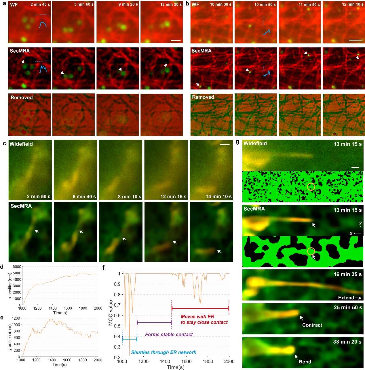
The researchers further proposed the Sectioning Multi-resolution Analysis (SecMRA) deconvolution algorithm, which introduces a newly designed biased sparse regularization that effectively improves performance in processing images with strong backgrounds. SecMRA-deconvolved widefield images achieve a similar sectioning effect to confocal microscopes, providing high-quality images while further reducing phototoxicity in live-cell imaging. The authors validated the effectiveness of this method across various imaging modalities, including widefield, Light-sheet, confocal, SoRa, SIM, STED, and Expansion microscopy.
The researchers demonstrated an example of using MRA deconvolution algorithm to support low light and low toxicity fluorescence imaging for studying cell activity. By using MRA deconvolution, researchers achieved long-term super-resolution imaging of microtubules and lysosomes co-labeled, observing the close relationship between lysosome movement and microtubule structure. The author also used a wide field microscope to achieve co-labeling imaging of mitochondria and endoplasmic reticulum around the nucleus for more than half an hour, observing the phenomenon of mitochondria rapidly extending, contracting, and finally binding to the endoplasmic reticulum.
In order to promote this technology, researchers have also proposed a solution to hyperparameter adjustment in regular deconvolution algorithms. By using the sparsity of the curvelet coefficient to estimate the noise intensity, automatic parameter determination has been achieved. The researchers developed user-friendly GUI software and open-source all the code and fluorescence image raw data for users and developers to use. The researchers hope that this work can provide a convenient deconvolution tool for biologists and microscopy researchers to use first-hand.
PhD student Hou Yiwei from the School of Future Technology, Peking University, is the first author of the paper. Professor Xi Peng and Dr. Li Meiqi from Peking University are the co-corresponding authors of the paper. The main contributors to the paper also include Fu Yunzhe, a PhD student at Peking University, and engineers Wang Wenyi and Ge Xichuan from Beijing Airy Precision Instrument Company. This work was supported by the National Key R&D Program of China and the National Natural Science Foundation of China.

Figure 2. SecMRA deconvolution enables long-term live cell imaging. a. b. Dual-color imaging of microtubules and lysosomes. SecMRA deconvolution effectively removes noise, out-of-focus background, and enhances resolution. c-g. SecMRA deconvolution achieves imaging of endoplasmic reticulum and mitochondrial interactions.


